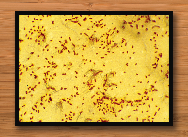
Photos of microscopic organisms enlarged and framed in new wing of science building

Dr. Jolie Stepaniak and lab associate Steve Schaar of HFC’s biology department are developing a plan for a series of photos of microscopic organisms to be enlarged, framed, and displayed in the new science wing in the science building (Bldg. J) on the main campus.
These photos are a result of the biology department purchasing two new microscopes with photographic capability. This will allow faculty members to take pictures for students, as well as take pictures of student-prepared specimens. Schaar was responsible for the purchase of these two microscopes. He also had the idea to enlarge the images of microscopic organisms and hang them in the new wing.
“The people involved in this project are excited to present the wonders of microscopic life to the students, staff, faculty, and administration of HFC,” said Schaar.
Variety of microscopic life
Images from a vast array of microscopic organisms will be included. Each photo will also have a description of the specimen, as well as information on sample preparation, magnification, microscopy, etc.
“We are planning on 8-10 microscopic images of living organisms,” said Stepaniak. “We haven't taken all the images yet, so the exact number will depend on how the images turn out. The images will represent a wide array of organisms at different magnifications. We plan to include an image of cardiac muscle, a tick, a plant specimen, a fungal specimen, and a nematode (roundworm).”